WELCOME TO MAMI
We are the only unit in SA to have been awarded the breast centre of excellence accreditation by the ACR. This is recognition of our dedication to excellence and our commitment to providing our patients with world class breast imaging.

Melrose Arch Mammography inc.
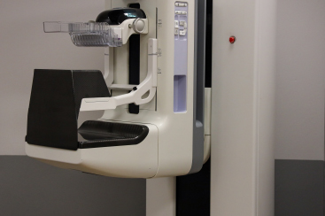
Melrose Arch Mammography inc. is the new face of Parklane Women’s Imaging Centre. The new practice has the same staff, state-of-the-art equipment and radiologists. But in a spacious, easily accessible, and beautiful new department. The Parklane practice was the first internationally accredited breast imaging unit in Africa. We participate in some of the leading breast cancer teams in the country, and are the only practice offering cancer cryoablation. At MAMI, you will have access to highly experienced doctors and the latest mammogram, ultrasound and biopsy equipment. We strive for medical excellence, but as importantly, our goal is to create a comfortable, caring environment for our patients.
MAMMOGRAPHY ACCREDITATION
The breast unit at Parklane Radiology (now Melrose Arch Mammography) is the first and only department in SA to achieve American College of Radiology (ACR) gold seal accreditation. Accreditation was awarded for mammography(2017), MRI (2018), Stereotactic Biopsy (2020) and Breast Ultrasound (2021). The department was designated a Breast Imaging Center of Excellence by the ACR in 2022.
The ACR gold seal of accreditation represents the highest level of image quality and patient safety. It is awarded only to facilities meeting ACR Practice Parameters and Technical Standards after a peer-review evaluation by board-certified physicians and medical physicists who are experts in the field. Image quality, personnel qualifications, adequacy of facility equipment, quality control procedures and quality assurance programs are assessed.
Diagnostic Radiologist & Managing Director
DR PETER SCHOUB
Parklane Radiology (PLR) was established in January 2012 when Drs Schoub and Said took over the hospital radiology department. At the time, the department was underutilized, and offered very limited services, mostly on obsolete equipment.
Dr Peter Schoub qualified as a doctor in 1998, and as a radiologist in 2007. For the last 15 years he has focused on breast radiology.
Dr Schoub joined Dr Harry Said at the Bagleyston Mammography Centre in 2011. The practice moved to the women’s imaging centre at Parklane Hospital in 2012.
The Parklane mammography unit became the first ACR (American College of Radiologists) unit in South Africa in 2017.
Dr Schoub works with several multidisciplinary breast cancer teams around Johannesburg. The largest, the Netcare Breast Care Centre of Excellence at Netcare Milpark Hospital, is fully accredited by the National Accreditation Program for Breast Centers (NAPBC), administered by the American College of Surgeons.
Within the context of the team, Dr Schoub works closely with surgeons and oncologists, assisting with diagnosis, staging, monitoring tumour response and presurgical localisation. He introduced cryoablation for breast cancers into the unit in 2020.
Dr Schoub obtained the European Diploma of Breast Imaging in 2018.
He is an honorary lecturer in the department of Radiology at the University of the Witwatersrand.


